A Tiny Buckle "Jack": A New Approach to Retinal Detachment Treatment – Successful FCB Surgery Performed by the Ophthalmology Department of Kunshan Hospital of Traditional Chinese Medicine
Release time: Jul 10,2025
FCB greatly improves the treatment experience of patients with retinal detachment. It is also worth mentioning that FCB implantation has low surgical risk and rapid recovery, making it more suitable for treating retinal detachment in patients with poor systemic conditions such as severe hypertension, diabetes, and heart disease.
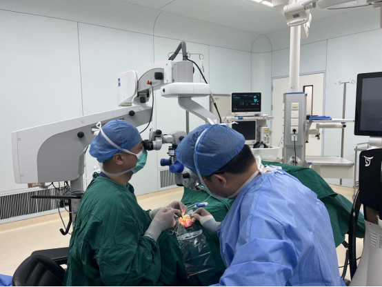
On May 12th, the Ophthalmology Department of Kunshan Hospital of Traditional Chinese Medicine successfully performed a Foldable Capsule Buckle (FCB) implantation. The team led by Director Yin Jian successfully implanted an FCB for a patient with complex rhegmatogenous retinal detachment, reattaching the detached retina. In fact, as early as November 19, 2024, Director Yin Jian had already used FCB to treat a patient with rhegmatogenous retinal detachment, effectively reattaching the retina and improving visual acuity. Kunshan Hospital of Traditional Chinese Medicine is also the first public hospital in Suzhou to carry out FCB surgery. As a new treatment option for rhegmatogenous retinal detachment, this surgical method marks that the ophthalmology department of our hospital has taken a further step towards minimally invasive, painless, and rapid treatment of rhegmatogenous retinal detachment.
Overview of the Condition
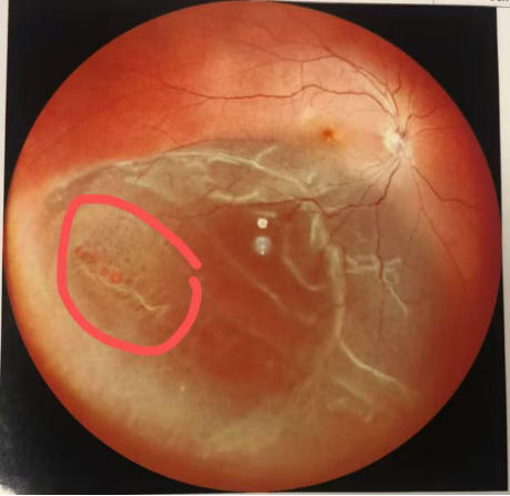
The patient was a 36-year-old female, admitted due to "right eye shadow occlusion for 8 days, vision loss with distortion for 2 days". Specialized examination: Visual acuity: VOD: 0.3 (corrected: -1.00D → 0.4-); VOS: 0.3 (corrected: -2.25D → 1.0). Intraocular pressure: right eye 17mmHg, left eye 16mmHg. The right eye showed no conjunctival hyperemia, clear cornea, normal depth of anterior chamber, Iyn (-), round pupil with a diameter of about 3.0mm, LR +, slightly increased lens density. Ocular examination: the optic disc had clear margins and normal color, with a C/D ratio of about 0.3. Multiple small round holes were seen in the peripheral retina of the inferotemporal area of the right eye; retinal detachment was present in the inferior and temporal areas, involving part of the retina below the macular area. B-ultrasound: mild vitreous opacity in the right eye, retinal detachment.

Traditional retinal detachment repair surgeries are divided into two types: internal and external approaches. The internal approach, namely vitrectomy, causes significant disturbance to intraocular tissues, with slow recovery, high cost, and requires the patient to maintain a prone position for 1 to 3 weeks after surgery. If silicone oil is used for tamponade, another surgery is needed to remove it after 1 to 3 months. The traditional external approach, namely scleral buckling, causes greater damage, slower recovery, and has a lower success rate. The patient hoped for a more minimally invasive treatment method with less impact on daily life and work. After thorough evaluation and communication, Director Yin's team performed FCB implantation for Mr. W.

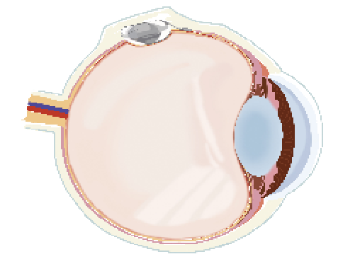
During the surgery, Director Yin made a small incision, implanted the folded buckle into a specific position between the conjunctiva and sclera of the patient's eyeball, and then injected saline to inflate the buckle. Like a jack, it compressed the scleral wall, thereby bringing the retinal pigment epithelium close to the retinal neuroepithelium at the hole, closing the hole, and promoting the reattachment of the detached retina. The surgery was successfully completed in 20 minutes.
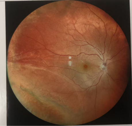
The patient adapted well after the surgery. However, due to the large number of holes and complex conditions, the medical staff successfully performed laser treatment to reattach the retina and close the holes. The patient was able to get out of bed and move freely soon after, was discharged smoothly, and only needed follow-up treatments in the outpatient department thereafter. The patient and their family expressed great satisfaction and sincere gratitude to Director Yin.

FCB Folded Capsule Buckle vs. Traditional External Surgery
FCB retinal reattachment surgery is a type of external approach surgery. In addition to the advantage of not disrupting the oxygen balance of the eye itself, it has many other benefits such as a smaller incision, shorter operation time, no retrobulbar anesthesia, and no muscle traction. Due to mild intraoperative reactions, rapid postoperative recovery, significantly reduced patient pain, and avoidance of many postoperative complications, FCB greatly improves the treatment experience of patients with retinal detachment. It is also worth mentioning that FCB implantation has low surgical risk and rapid recovery, making it more suitable for treating retinal detachment in patients with poor systemic conditions such as severe hypertension, diabetes, and heart disease.


