Tangshan Aier Eye Hospital Successfully Performs FCB Surgery for Ultra-Minimally Invasive and Rapid Treatment of Retinal Detachment
Release time: Aug 20,2025
Tangshan Aier Eye Hospital Successfully Performs FCB Surgery for Ultra-Minimally Invasive and Rapid Treatment of Retinal Detachment
Tangshan Aier Eye Hospital has successfully carried out FCB surgery, providing ultra-minimally invasive and rapid treatment for retinal detachment. On August 16th, Professor Liu Junhui from Tangshan Aier Eye Hospital successfully performed a Foldable Capsular Buckle (FCB) implantation surgery. He smoothly implanted an FCB for a patient with rhegmatogenous retinal detachment, achieving successful reattachment of the patient’s detached retina. As a new surgical method for the treatment of rhegmatogenous retinal detachment, FCB implantation is a special type of scleral buckle surgery characterized by minimal invasiveness and fewer complications. After the surgery, the patient can maintain a free body position, which greatly improves the overall surgical experience and provides a safer and more efficient surgical option for patients.
Patient Profile
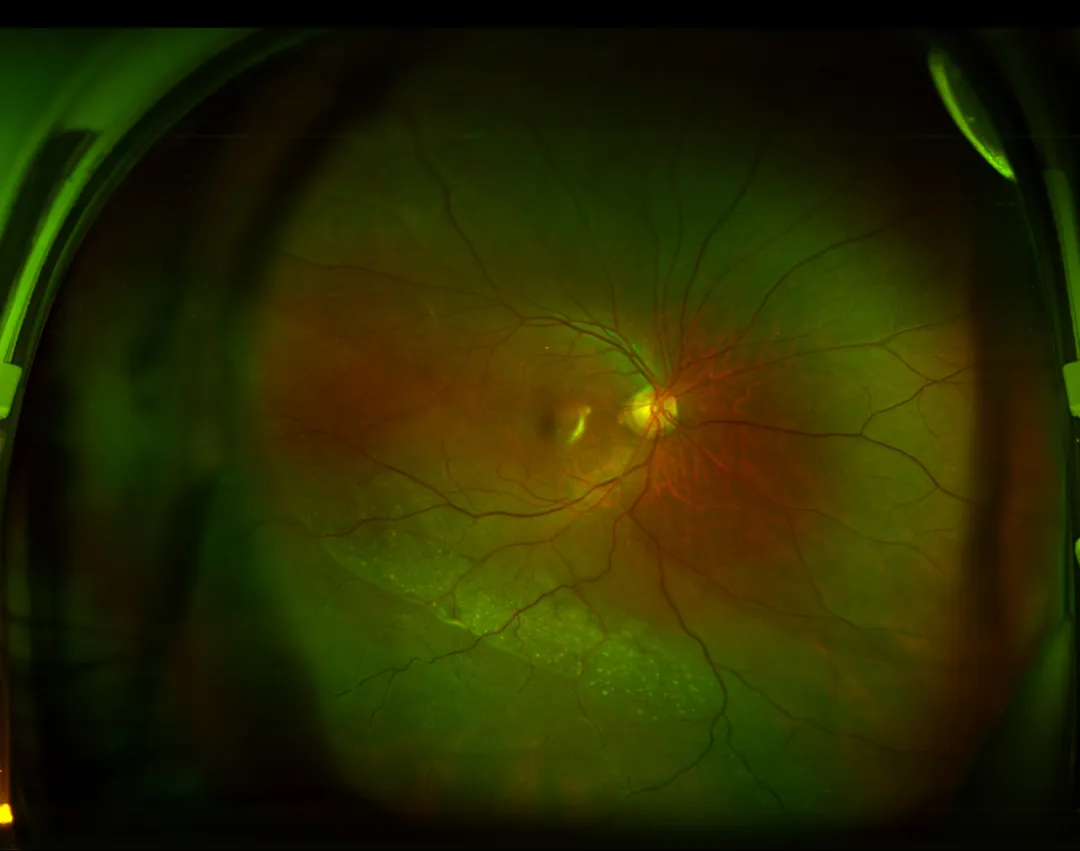
The patient was a 61-year-old male, admitted to the hospital due to "black shadow obstruction in the upper part of the right eye for 3 days". Admission examination results: - Visual acuity (uncorrected): Right eye 1.0, Left eye 1.2 - Intraocular pressure (IOP): Right eye 17mmHg, Left eye 17mmHg - Fundus of the right eye: The optic disc had a clear border and normal color; there was a grayish-white retinal elevation in the inferotemporal 4:00-10:00 area, involving the inferotemporal macular region; yellow-white and grayish-white particles were attached under the detached retina; a hole was found in the peripheral retina at the 8:30 position, with eversion of the hole margin; clustered vitreous and small hemorrhage were observed at the edge of the hole; no foveal light reflex was seen. (Image omitted)

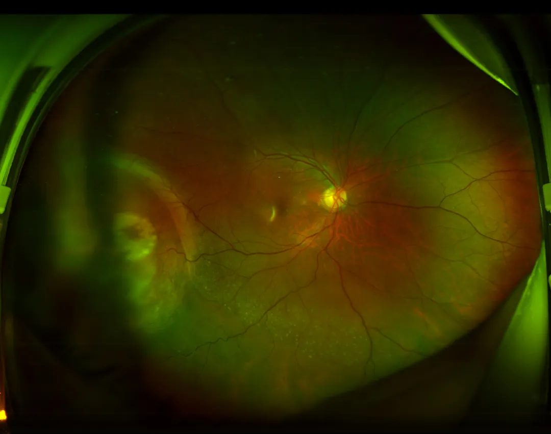
Given that the patient had a single retinal hole (located around the equator, relatively fresh) and was elderly, the patient’s family hoped for a painless and minimally invasive treatment as much as possible. Based on the patient’s condition, Professor Liu Junhui formulated an appropriate treatment plan. After full communication with the patient and his family, it was decided to perform the latest minimally invasive retinal reattachment surgery for him — Foldable Capsular Buckle (FCB) retinal reattachment surgery. On the 2nd day after admission, Professor Liu’s team performed FCB implantation on the patient’s right eye. The surgery took only more than 10 minutes, and successful retinal reattachment was achieved. On the first day after the surgery, retinal laser photocoagulation was performed on the right eye. Postoperative examination results: - Visual acuity (uncorrected): Right eye 0.8, Left eye 1.0 - Intraocular pressure (IOP): Right eye 19mmHg, Left eye 16mmHg - Other findings of the right eye: Mild conjunctival hyperemia; the surgical incision was well closed with sutures in place; the cornea was transparent; fundus examination showed a clear optic disc with normal color, a moderate-to-high compression ridge, the retinal hole located on the ridge, good response of the surrounding laser spots, and a small amount of subretinal fluid in the lower area. (Postoperative examination image omitted)

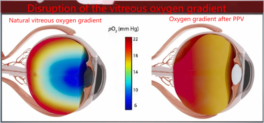
## FCB: Ultra-Minimally Invasive and Rapid Treatment for Retinal Detachment ### 01 Internal Surgery vs. External Surgery - **Internal Surgery**: Also known as pars plana vitrectomy (PPV). It causes severe and irreversible damage, has a slow recovery process and high cost, and requires the patient to rest in a prone position for 2 to 3 weeks after surgery. If silicone oil tamponade is used, another surgery is needed to remove the silicone oil after 1 to 3 months. Vitrectomy destroys the oxygen gradient of the vitreous, which may lead to irreversible conditions such as cataracts and optic nerve damage.

**External Surgery**: Recommended by more doctors. It does not require entering the eye, thus ensuring that the eye’s own oxygen balance is not disrupted. The traditional external surgery is external scleral buckling, which causes less damage than internal surgery but has a lower success rate. Once successful, no repeated surgeries are needed, reducing the patient’s suffering.
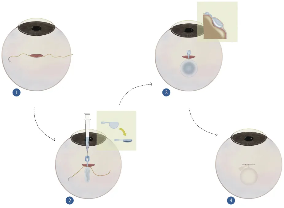
### 02 Foldable Capsular Buckle (FCB) Surgery Foldable Capsular Buckle (FCB) retinal reattachment surgery is a type of external surgery. Compared with traditional external retinal surgery, FCB has the following advantages: - Shortened operation time (reduced from 50 minutes to 10 minutes) - No retrobulbar anesthesia - No muscle traction - No scleral fluid drainage - No intraoperative localization - No hole cryotherapy The doctor only needs to implant the folded balloon into the outer wall of the eye at the detachment site, then inject normal saline to inflate the balloon. Like a jack, the balloon "pushes back" the detached retina. This surgery enables fast postoperative recovery, greatly reduces the patient’s pain, avoids many postoperative complications, and significantly enhances the treatment experience of patients with retinal detachment (abbreviated as "wang Tuo" in Chinese). The surgery adopts a 3D reconstruction calculation method to accurately determine the location of the retinal hole, which can effectively improve the accuracy of hole reattachment and the overall retinal reattachment rate.

Doctor’s Profile

Professor Liu Junhui - Associate Chief Physician - Director of the Fundus Disease Department, Associate Chief Physician, Master’s Degree - Has been engaged in clinical ophthalmology work for more than 20 years - Serves as a Member of the Fundus Disease Group of Aier Eye Hospital (Hebei Region) - Member of the Ophthalmology Special Committee of the China Population Culture Promotion Association - Member of the Eye Disease Prevention, Treatment and Rehabilitation Professional Committee of Hebei Community Rehabilitation Medical Association
The surgery not only averted the risk of eyeball atrophy but also preserved his precious visual function.
12/30
Two days later, laser photocoagulation was performed on the retinal hole in the right eye. Postoperative examination showed the retinal hole was located on the buckle ridge with good adhesion. The patient and her family were satisfied with the postoperative outcome and expressed their gratitude to Director Li's team!
12/24


