

Patient-centered, physician-partner, employee-based
The 3rd Session of International Ophthalmic Innovative Surgery Live Broadcast
Release time: Sep 08,2025
Series Events of China-Europe (Germany) Ophthalmology R&D and Innovation Center: The 3rd Session of International Ophthalmic Innovative Surgery Live Broadcast - "Ultra-Minimally Invasive Treatment of RRD with FCB" Launches!
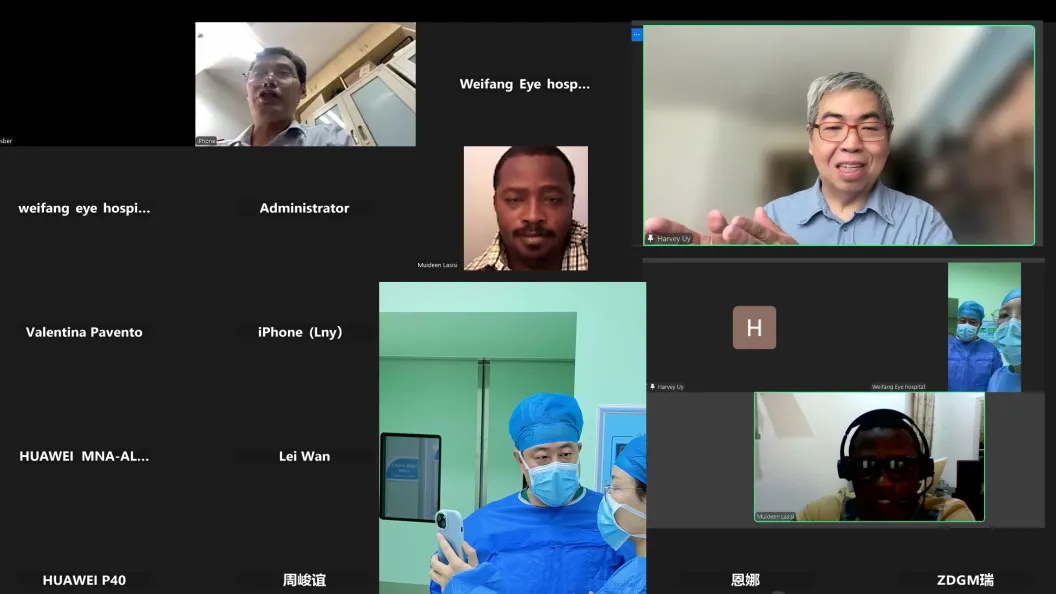
On September 3, the 3rd session of the International Ophthalmic Innovative Surgery Live Broadcast, themed "Ultra-Minimally Invasive Treatment of RRD with FCB", under the series events of China-Europe (Germany) Ophthalmology R&D and Innovation Center, was successfully launched. This live broadcast specially invited Professor Zhang Jie, President and Party Branch Secretary of Weifang Eye Hospital, as the surgical guest, and Prof. Harvey Uy M.D., Ph.D. from the Philippines as the overseas guest representative. Prof. Harvey Uy currently serves as the Medical Director of Peregrine Eye and Laser Institute and is also the President of this year's Asia-Pacific Vitreoretina Society (APVRS 2025) Congress in Manila. The two experts jointly demonstrated the application of Foldable Capsular Buckle (FCB) implantation in the treatment of Rhegmatogenous Retinal Detachment (RRD) to more than 100 ophthalmologists and specialists at home and abroad. Ophthalmology experts from 15 countries, including Italy, Spain, Singapore, Qatar, Peru, Turkey, the Philippines, and Indonesia, watched and learned online simultaneously. They jointly discussed how to improve the quality and efficiency of retinal disease diagnosis and treatment through technological innovation, aiming to promote the global level of retinal disease diagnosis and treatment to a new height.
Breakthrough Achieved in Ultra-Minimally Invasive Surgery for Retinal Detachment
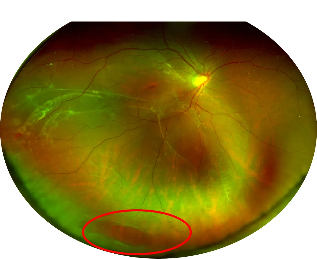
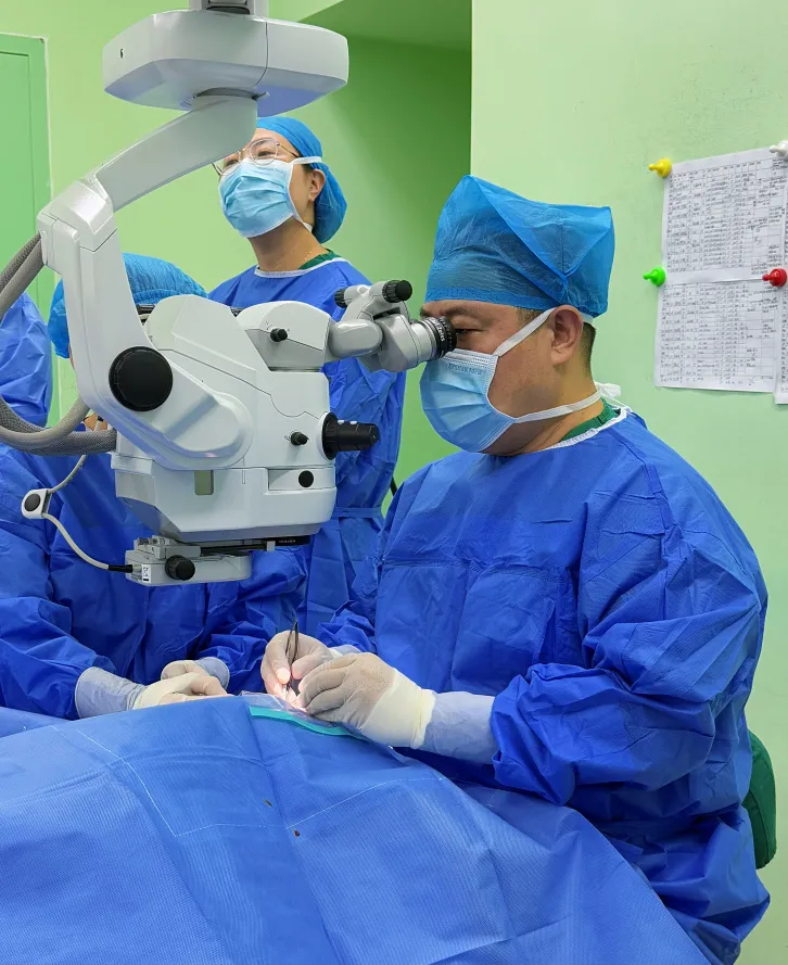
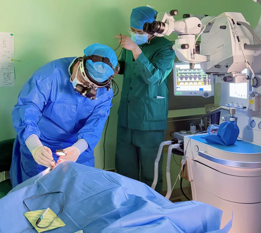
At the beginning of the live broadcast, Professor Zhang Jie briefly introduced the patient's condition. The patient was a 20-year-old male, who was admitted to the hospital with the main complaint of blurred vision and a sense of obstruction in the right eye for 3 months. Examination results showed: the visual acuity of the right eye was 0.05, and the corrected visual acuity was 0.1; the retina at the 4:00-6:00-9:00 positions of the right eye fundus showed a grayish-blue detachment, involving the macula; an elliptical tear of approximately 3 PD (papillary diameter) in size was observed in the peripheral retina at the 6:00 position. The condition was relatively severe.
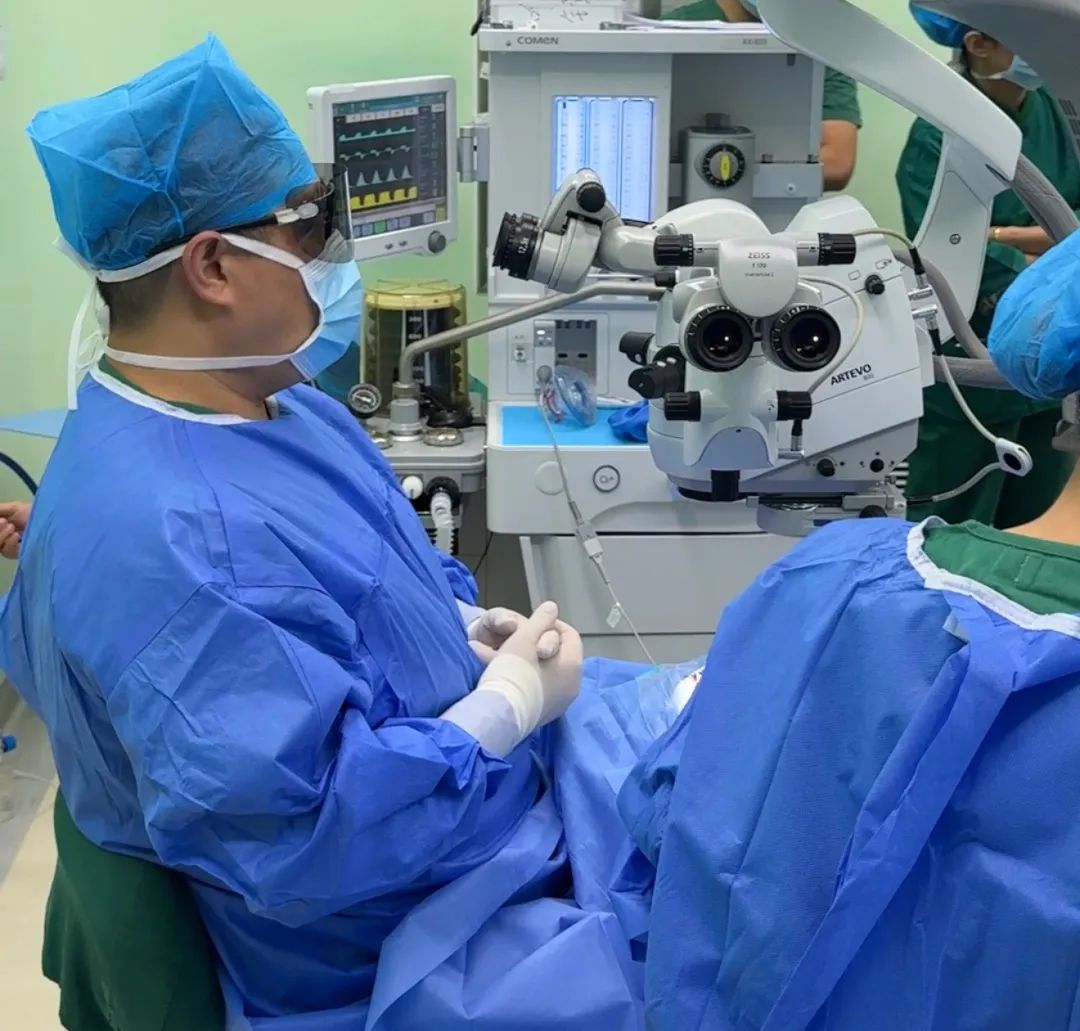
Subsequently, in the live surgery session, relying on his exquisite surgical skills and in-depth understanding of FCB, Professor Zhang Jie implemented a highly innovative personalized surgical plan. Through the "four 'no' operations" - no retrobulbar anesthesia, no muscle traction, no intraoperative localization, and no tear cryotherapy - he successfully treated the retinal detachment patient with FCB-based ultra-minimally invasive surgery. Professor Zhang's superb skills not only ensured the smooth completion of the surgery but also significantly shortened the operation time: the traditional external scleral buckling surgery usually takes 50 minutes, while the FCB surgery time can be effectively reduced to only 10 minutes. During the surgery, the patient reported no special discomfort, the operation went smoothly, and after the surgery, the retinal tear in the patient's right eye was ideally closed with good therapeutic effect, which satisfied the patient.
The surgery performed by Professor Zhang Jie is a perfect demonstration of ultra-minimally invasive external treatment of RRD using Foldable Capsular Buckle (FCB). Prof. Harvey Uy M.D., Ph.D. highly commented on the surgical process, praising Professor Zhang for his "exquisite skills" and fully affirming the outstanding performance of this innovative surgical method in terms of accuracy and efficiency. This surgery not only demonstrates the cutting-edge level of ophthalmic surgical technology but also marks that ophthalmic clinical treatment is accelerating its transformation towards the direction of "minimally invasive and precise", providing a brand-new idea and reference for ophthalmologists worldwide in the field of retinal detachment treatment.
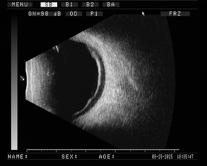
After the surgery, President Zhang used an indirect ophthalmoscope to check the tamponade position of the patient's retinal tear. He confirmed that the tamponade position was good and the preoperative localization was relatively accurate, thus verifying the successful completion of the surgery.
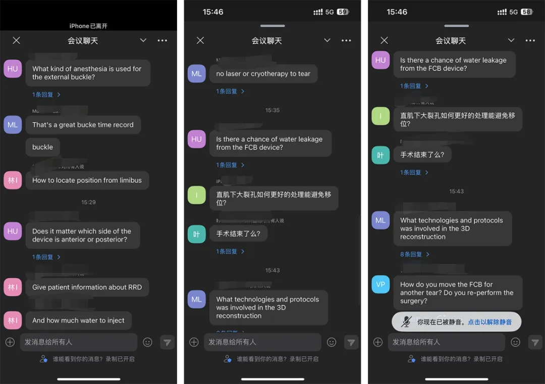
Finally, Professor Zhang, Professor Harvey, and other experts at home and abroad online conducted in-depth and lively discussions on topics such as FCB indication selection, operation standards, patient comfort, inclusion criteria, and surgical operations, sharing their rich clinical experience and unique insights.

The ultra-minimally invasive surgery demonstrated in this live broadcast has redefined the treatment method for retinal detachment. This surgery marks a paradigm shift in ophthalmic intervention towards "minimally invasive and precise". Professor Harvey particularly affirmed the low invasiveness of this surgical method - this feature enables patients to recover faster after surgery, reduces the occurrence of postoperative complications, and significantly improves the patient's medical experience. In addition, the discussion on FCB costs and global accessibility has also become a focus, highlighting its potential to go beyond China's domestic market and innovate global medical care.
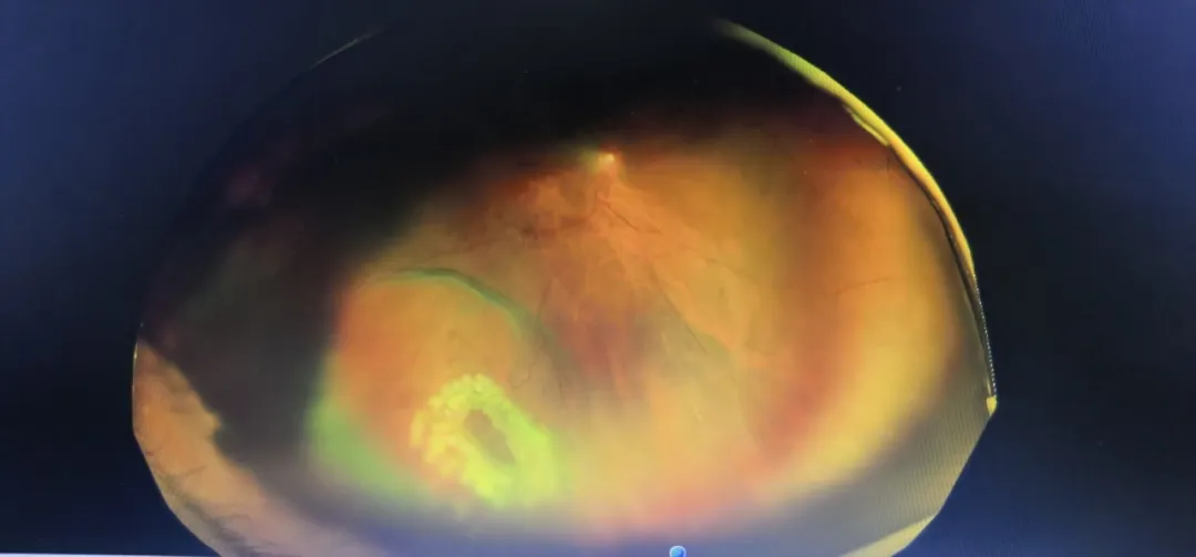
It is worth mentioning that the fundus examination results of the patient after laser treatment on the second day post-operation showed that the FCB tamponade effect was good and the retinal reattachment was stable. This further verifies the success of the surgery and the reliability of FCB technology. This result not only brings hope for the patient to regain clear vision but also provides strong clinical evidence for ophthalmologists worldwide in the treatment of retinal detachment.
3D Reconstruction of FCB Successfully Published in Clinical Ophthalmology
This research not only shortens the learning curve and lowers the technical threshold of FCB surgery, but also lays a solid foundation for its standardized promotion in primary hospitals and among special populations (such as children, pregnant women, and highly myopic patients), truly realizing a minimally invasive, precise and reproducible clinical pathway.
03/31
The APAO 2026 congress served as an academic feast and international exchange platform for the ophthalmology community. As a globally innovative technology, the Foldable Capsular Buckle (FCB) sparked considerable discussion at APAO 2026, fully reflecting the technology's visibility and research depth
02/10
On the day of the surgery, Professor Hou Lei and Professor Chen Zhongping worked closely together, leveraging their exceptional medical skills and seamless collaboration to successfully complete Qinghai Province's first FCB procedure.
02/05

R&D Center
Media
Cases
Search for product information you are interested in
CONTACT US
+8618960050093
Building11,No.6,Nanjiang 2nd Road, Zhujiang Street, Nansha District,Guangzhou


©COPYRIGHT 2024 Guangzhou Vesber Biotechnology Co., Ltd. | SEO Tags Privacy Policy