

Patient-centered, physician-partner, employee-based
Tiny Capsule, Big Miracle! Ningxiang Aier Eye Hospital Successfully Performs Foldable Capsular Buckle (FCB) Implantation
Release time: May 20,2025
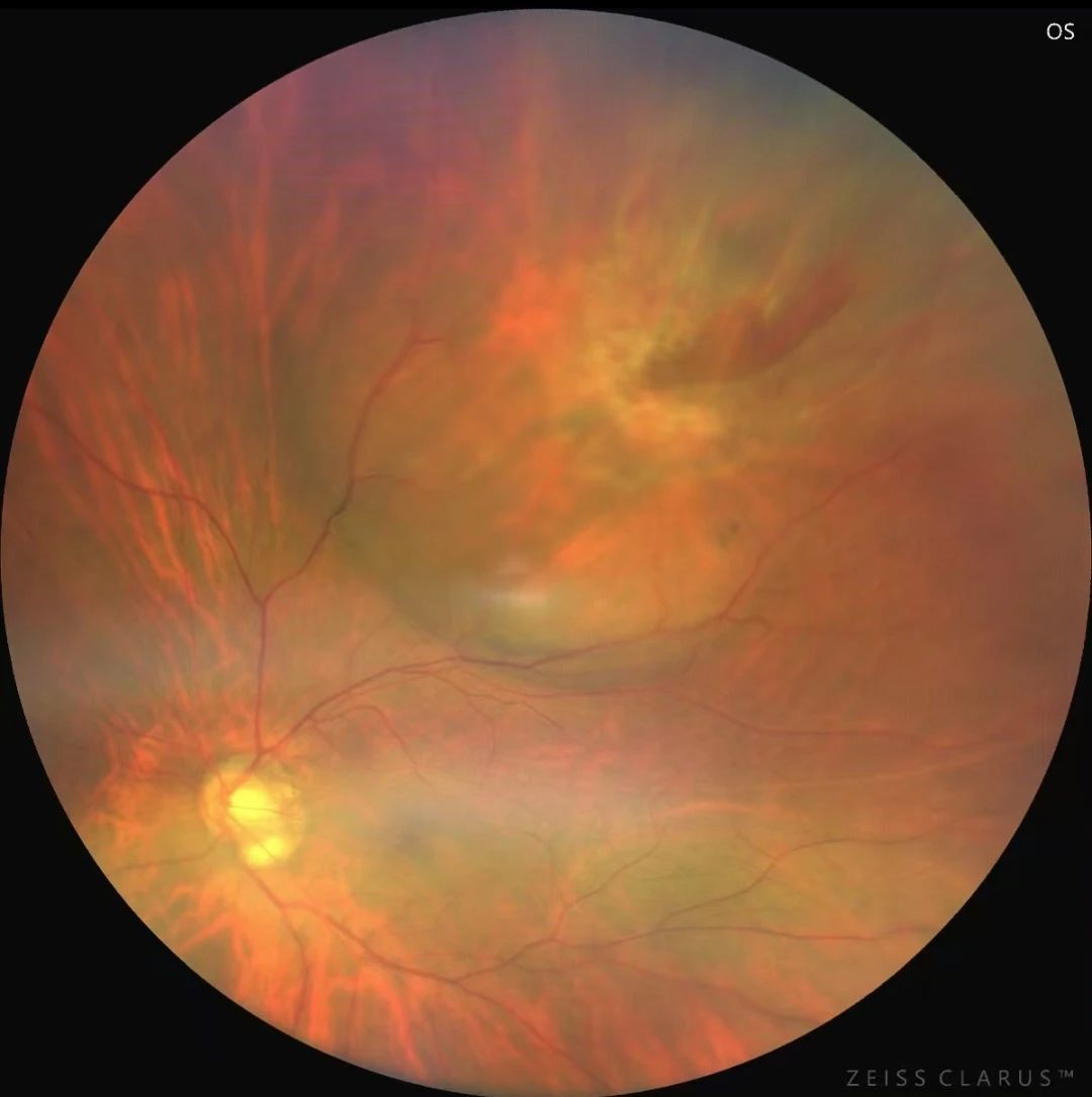
As a new treatment for rhegmatogenous retinal detachment, this procedure marks a step forward for the hospital’s ophthalmic fundus disease department toward minimally invasive, painless, and efficient therapy.
Case Overview
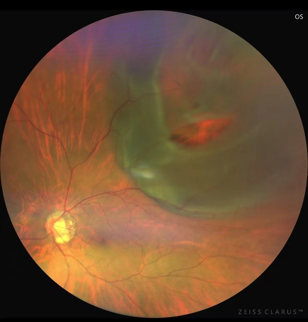
Treatment Plan
Key Innovations
- 3D Reconstruction Technology: Precisely locates the tear site to enhance reattachment accuracy and success rate.
- Minimally Invasive Advantages: Smaller incisions reduce postoperative complications, and the removable implant avoids long-term intraocular foreign bodies.
- Shorter Operation Time: Safer for patients with systemic conditions (e.g., pregnancy, diabetes, hypertension) by minimizing the risk of cardio-ocular reflex during surgery.

R&D Center
Media
Cases
Search for product information you are interested in
CONTACT US
+8618960050093
Building11,No.6,Nanjiang 2nd Road, Zhujiang Street, Nansha District,Guangzhou


©COPYRIGHT 2024 Guangzhou Vesber Biotechnology Co., Ltd. | SEO Tags Privacy Policy
License