Jinan Mingshui Eye Hospital has successfully carried out the FCB surgery, providing a new treatment method for patients with retinal detachment due to large breaks.
Release time: Apr 25,2025
The FCB implantation does not change the intraocular environment, greatly shortens the operation time, and allows the patient to lie in a free position after the operation, enhancing the surgical experience.
On March 20, President Wang Donglin of Jinan Mingshui Eye Hospital successfully performed the implantation of the foldable Capsular Buckle (FCB). He successfully implanted the FCB for a patient with rhegmatogenous retinal detachment due to a large break, repositioned the detached retina, and improved the visual acuity. The FCB implantation does not change the intraocular environment, greatly shortens the operation time, and allows the patient to lie in a free position after the operation, enhancing the surgical experience.
Overview of the Condition
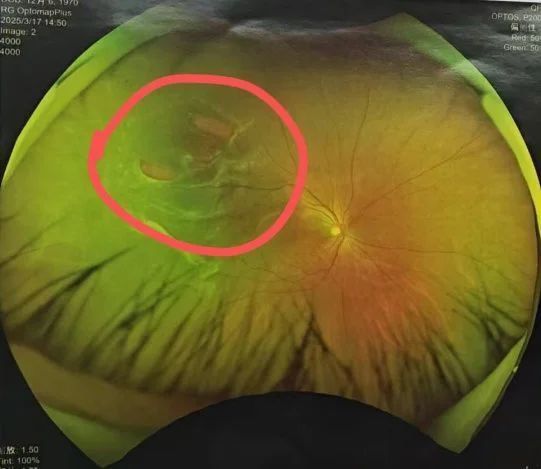
The patient is a middle-aged woman who was admitted to the hospital with the chief complaint of "blurred vision in the right eye for half a month". The patient stated that she developed blurred vision in the right eye accompanied by a sense of visual obstruction on the nasal side after "working overtime continuously for half a month", and the symptoms gradually worsened. To seek diagnosis and treatment, she visited a nearby hospital one day ago and was diagnosed with "rhegmatogenous retinal detachment (right eye)", but no treatment was given. In order to have a more definite diagnosis and treatment, she came to Jinan Mingshui Eye Hospital. After outpatient examination, she was diagnosed with "rhegmatogenous retinal detachment (right eye)", and a right eye retinal reattachment surgery was recommended. The team led by Professor Wang Donglin conducted a specialized examination of her vision: the visual acuity of the right eye was 0.3 and that of the left eye was 0.8; the intraocular pressure: 14 mmHg in the right eye and 15 mmHg in the left eye; the fundus of the right eye: the optic disc had a clear boundary and normal color, the large arteriovenous vessels were normal, the retina was elevated from about 7 o'clock to 11 o'clock, involving the macular area. A horseshoe-shaped tear was formed around 10 o'clock, with a size of about 7 PD, and the reflection of the fovea centralis of the macula was not clear.
Considering the patient's condition, Professor Wang took into account that the onset time of the right eye was half a month and the break was still fresh, especially as the break was as large as 7 PD. If the traditional external approach surgery was adopted, the success rate would be low. Therefore, in the past, many doctors would perform an internal approach vitrectomy, but there were many complications after vitrectomy. The patient hoped for a minimally invasive and effective treatment. Therefore, Professor Wang formulated an appropriate treatment plan. After fully communicating with the patient and her family, he carried out the latest minimally invasive retinal reattachment surgery for her - the implantation of the foldable capsular buckle(FCB).
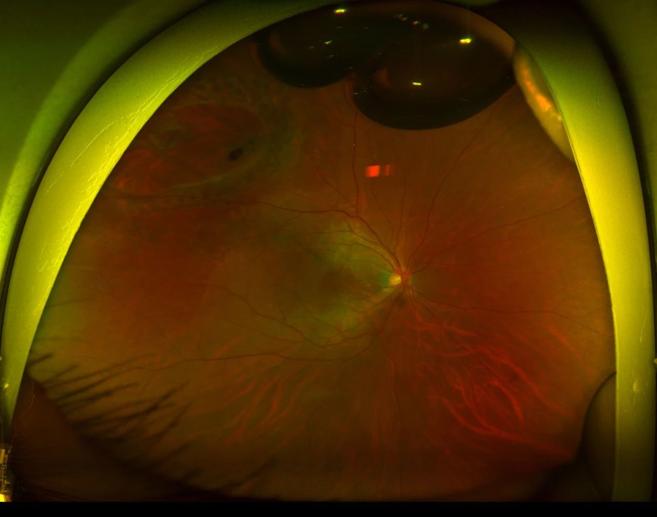
The day after the patient was admitted to the hospital, local anesthesia was administered, and the FCB in the right eye was combined with the injection of C3F8 into the vitreous cavity. After the operation, laser photocoagulation of the retinal tear in the right eye was carried out, along with anti-inflammatory treatment, prevention of infection, and pupil dilation for symptomatic treatment. After the operation, the visual acuity of the right eye improved, and the retina became flat. The patient was satisfied with the postoperative effect and expressed deep gratitude to the medical team! The full efforts of both the doctor and the patient enabled the patient to avoid the fate of vitrectomy, which also gave the patient an opportunity.
The successful implementation of Chenzhou Aier Ophthalmology Hospital's first FCB surgery not only brought light to Xiao L but also marked that the hospital has stepped into a new stage in the field of minimally invasive treatment for retinal detachment. It provides a higher-quality and more precise treatment option for patients with retinal detachment in southern Hunan, especially adolescent patients.